
Current science about DNA methylation
Main difference between genetics and epigenetics
Genetics and epigenetics look at different parts of our biology. Genetic information is focused on the sequence (the order of nucleotides) of the adenine (A), cytosine (C), guanine (G), and thymine (T) molecules that make up the base pairs in an individual’s DNA, including the presence or absence of specific mutations.
By contrast, epigenetic information involves the presence or absence of chemical modifications that may affect the processes of gene expression and translation into proteins without altering the DNA sequence itself.

Cytosine - phosphate - Guanine nucleotide sites (CpG)
The CpG sites or CG sites are regions of DNA where a cytosine nucleotide is followed by a guanine nucleotide in the linear sequence of bases along its 5' → 3' direction. Phosphate links any two nucleosides together in DNA. The CpG notation is used to distinguish this single-stranded linear sequence from the CG base-pairing of cytosine and guanine for double-stranded sequences. CpG sites occur with high frequency in genomic regions called CpG islands (or CG islands). Cytosines in CpG dinucleotides can be methylated to form 5-methylcytosines. Enzymes that add a methyl group are called DNA methyltransferases (DNMTs).

DNA Methyltransferases (DNMTs)
DNA methyltransferases (DNMTs), including DNMT1, DNMT3a and DNMT3b, catalyze the methylation of CpG dinucleotides by addition of a methyl group from S-adenosyl-L-methionine to the 5’ carbon position of cytosine. DNMTs are responsible for setting and maintaining DNA methylation patterns. DNMT3a and DNMT3b methylate previously unmethylated CpG sequences and are classified as de novo methyltransferases. In contrast, DNMT1, the most prevalent methyltransferase in mammals, ensures the maintenance of methylation tags onto new DNA strands during replication. Diseased cells such as cancer cells may be different in that DNMT1 alone is not responsible for maintaining normal gene hypermethylation (an increase in global DNA methylation) and both DNMTs 1 and 3b may cooperate for this function.
DNMT3a and DNMT3b are DNA modifying proteins mostly involved in the methylation of CpG sequences during embryogenesis. Different from DNMT1, DNMT3 enzymes do not target hemimethylated DNA. Expression of DNTM3 enzymes is induced at embryonic stages and downregulated in differentiated cells.
Methylation of DNA by DNMTs leads to gene silencing. This epigenetic mark is involved in normal processes such as aging and cell differentiation, but is also thought to play a role in the development of diseases such as cancer, Alzheimer's disease, and many others.

What is DNA methylation?
DNA methylation is a biological process by which methyl groups are added to the DNA, specifically cytosine molecule. Methylation therefore can change the activity of a DNA segment without changing the sequence.
As mentioned above, when located in a gene promoter, DNA methylation typically acts to repress gene expression (gene silencing).
Methylation the cytosine within a gene can change its expression, a mechanism that is part of a larger field of epigenetics. In humans, about 70% of promoters located near the transcription start site of a gene (proximal promoters) contain a CpG island.

DNA demethylation
DNA demethylation is the removal of a methyl group from DNA. This mechanism is equally as important and coupled with DNA methylation. Hypomethylation of CpG islands in promoters results in overexpression of the genes or gene sets affected.
The demethylation process is necessary for epigenetic reprogramming of genes and is also directly involved in many important disease mechanisms such as tumor progression. Demethylation of DNA can either be passive or active, or a combination of both. Passive DNA demethylation usually takes place on newly synthesized DNA strands via DNMT1 during replication rounds. Active DNA demethylation mainly occurs by the removal of 5-methylcytosine via the sequential modification of cytosine bases that have been converted by TET enzyme-mediated oxidation.

DNA repair genes with hyper/hypo-methylated promoters in cancers
DNA repair genes are frequently repressed in cancers due to hypermethylation of CpG islands within their promoters.
In head and neck squamous cell carcinomas at least 15 DNA repair genes have frequently hypermethylated promoters; these genes are XRCC1, MLH3, PMS1, RAD51B, XRCC3, RAD54B, BRCA1, SHFM1, GEN1, FANCE, FAAP20, SPRTN, SETMAR, HUS1, and PER1. About seventeen types of cancer are frequently deficient in one or more DNA repair genes due to hypermethylation of their promoters. As an example, promoter hypermethylation of the DNA repair gene MGMT occurs in 93% of bladder cancers, 88% of stomach cancers, 74% of thyroid cancers, 40%-90% of colorectal cancers and 50% of brain cancers. Promoter hypermethylation of LIG4 occurs in 82% of colorectal cancers. Promoter hypermethylation of NEIL1 occurs in 62% of head and neck cancers and in 42% of non-small-cell lung cancers. Promoter hypermethylation of ATM occurs in 47% of non-small-cell lung cancers. Promoter hypermethylation of MLH1 occurs in 48% of non-small-cell lung cancer squamous cell carcinomas. Promoter hypermethylation of FANCB occurs in 46% of head and neck cancers. On the other hand, the promoters of two genes, PARP1 and FEN1, were hypomethylated and these genes were overexpressed in numerous cancers. PARP1 and FEN1 are essential genes in the error-prone and mutagenic DNA repair pathway microhomology-mediated end joining. If this pathway is overexpressed the excess mutations it causes can lead to cancer. PARP1 is overexpressed in tyrosine kinase-activated leukemias, in neuroblastoma, in testicular and other germ cell tumors, and in Ewing's sarcoma, FEN1 is overexpressed in the majority of cancers of the breast, prostate, stomach, neuroblastomas, pancreatic, and lung.
DNA damage appears to be the primary underlying cause of cancer. If accurate DNA repair is deficient, DNA damages tend to accumulate. Such excess DNA damage can increase mutational errors during DNA replication due to error-prone translesion synthesis. Excess DNA damage can also increase epigenetic alterations due to errors during DNA repair. Such mutations and epigenetic alterations can give rise to cancer (see malignant neoplasms). Thus, CpG island hyper/hypo-methylation in the promoters of DNA repair genes are likely central to progression to cancer.
Methylation of CpG sites with age
Since age has a strong effect on DNA methylation levels on tens of thousands of CpG sites, one can define a highly accurate biological clock (referred to as epigenetic clock or DNA methylation age) in humans and chimpanzees.

Unmethylated sites
Unmethylated CpG dinucleotide sites can be detected by Toll-like receptor 9 (TLR 9) on plasmacytoid dendritic cells, monocytes, natural killer (NK) cells, and B cells in humans. This is used to detect intracellular viral infection.
How is DNA methylation analyzed? Bisulfite conversion process.
For epigenetic diagnostics it is important to provide highest precision for the DNA methylation level at individual CpG sides. To this end, DNA needs to be isolated, and bisulfite treated to convert unmethylated cytosines into thymidine bases, whereas the 5-methylcytosine residues remain protected from this conversion. The thymidine to cytosine ratio is therefore indicative for DNA methylation of specific CpG sites. The results are then integrated into mathematical models to combine the predictive power of several CpG sites.
A key limitation of using a bisulfite sequencing method is the chance of incomplete conversion. The bisulfite conversion process also raises issues as the necessary conditions may damage the DNA. High temperatures and long incubation times can lead to approximately 90% DNA degradation.
Common DNA methylation methods
Common global DNA methylation methods include HPLC, LC-MS/MS, LUMA, and M.SssI acceptance assays. HPLC uses enzymatic hydrolysis of genomic DNA and typically requires large amounts of sample DNA. Furthermore, optimization of the procedure requires extensive expertise. LC-MS/MS is considered the "gold standard" for quantification of global DNA methylation and similarly digests and separates deoxyribonucleotides, but analysis by mass spectrometry requires highly specialized knowledge. LUMA is a method that relies on cleaving the DNA with methylation-sensitive restriction enzymes followed by pyrosequencing. It isn't conclusive as to whether or not the results from this method accurately represent global DNA methylation. Lastly, the M.Sssl acceptance assay requires radioactivity for analysis and utilizes CpG DNA methyltransferase M.SssI and methyl donor S-adenosylmethionine.
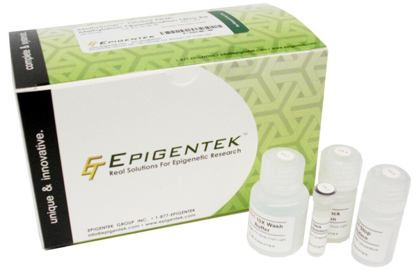
Directly quantifying total 5-mC for an accurate measurement of global DNA methylation can be accomplished in under 4 hours using the MethylFlash technique, a simple ELISA-like method.
Future for the field
CRISPR/Cas9 technology is an important advance in the field of genetics and its editing methods may be applied to the epigenome. Primarily, this may be used to give greater clarification regarding the relationship between methylation and cell behavior and function; information that may later be translated to the clinic.